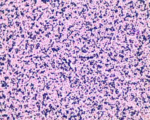
200以上 e coli gram stain 400x 210005-E coli gram stain 400x
Total mag = 1000X) mixture = Gram positive and Gram negative organsims (Staphylococcus aureus and E coli) Gram negative organisms Ecoli Gram positive organisms Gram coccus Gram Bacillus Gram positive rod for comparison = yeast (unstained) at 400XClinical Significance E coli colonizes the large intestine of humans It is the most common pathogen of urinary tract infections and is associated with pneumonia, bacteremia (typically secondary to urinary tract infections), endocarditis, meningitis (typically in neonates), hepatic abscesses, peritonitis, endophthalmitisGram Stain 5 Review Stains Endospore, Capsule & Bacteria E coli Must observe under 400x Very small & motile Staph Gram positive E coli Gram negative cocci in clusters rods (no arrangement) Refer to Lab Manual for directions Instructor to

Bacterial Morphotypes In Sputum Gram Stain 100 Oil Immersion Field Download Scientific Diagram
E coli gram stain 400x
E coli gram stain 400x-A) The smear is too thick B) The slide is washed with ethyl alcohol for 30 secs during the staining technique C) The slide is heatfixed until the smear turns yellow/brown D) The bacterial culture is very old E) Cold water is used to wash the slide during the stainingTotal mag = 1000X) mixture = Gram positive and Gram negative organsims (Staphylococcus aureus and E coli) Gram negative organisms Ecoli Gram positive organisms Gram coccus Gram Bacillus Gram positive rod for comparison = yeast (unstained) at 400X


Biol 2
E Coli E Coli under the microscope at 400x E Coli (Escherichia Coli) is a gramnegative, rodshaped bacterium Most E Coli strains are harmless, but some serotypes can cause food poisoning in their hosts The harmless strains are part of the normal flora of the gut Learn more about E Coli here Helicobacter PyloriUsing Gram Staining (Lab 4L) (Cat # BE2) and in sharp focus on 100X before going to 400X, and center and focused on 400X before adding the immersion oil and going to 1000X Students often use too much light when using a microscope Decreasing the diaphragm E coli cells are GramWhen a report says "rare gram negative rod," it
The key difference between E coli and Pseudomonas aeruginosa is that E coli is a facultative anaerobic bacterial species that belongs to family Enterobacteriaceae and genus Escherichia, while P aeruginosa is an aerobic bacterial species that belongs to family Pseudomonadadaceae and genus Pseudomonas Both E coli and Pseudomonas aeruginosa are gramnegative, rodshaped and motile bacteriaEscherichia coli (/ ˌ ɛ ʃ ə ˈ r ɪ k i ə ˈ k oʊ l aɪ /), also known as E coli (/ ˌ iː ˈ k oʊ l aɪ /), is a Gramnegative, facultative anaerobic, rodshaped, coliform bacterium of the genus Escherichia that is commonly found in the lower intestine of warmblooded organisms (endotherms) Most E coli strains are harmless, but some serotypes (EPEC, ETEC etc) can cause serious foodEscherichia Coli (400x) Escherichia coli (E Coli) is one of many species of bacteria that lives in the lower intestines of mammals In the large intestine of mammals, E coli assists in waste processing, vitamin K production and food absorption E coli causes illness in humans
Oct 28, 15 Acid fast stain of Echerichia coli 400x on a compound light micrscope Echerichia coli is a gram negative, non spore forming capsulated bacteriaGram Stain (all 100X oil immersion objective plus zoom on camera) (eyepiece = 10X;Escherichia Coli (400x) Escherichia coli (E Coli) is one of many species of bacteria that lives in the lower intestines of mammals In the large intestine of mammals, E coli assists in waste processing, vitamin K production and food absorption


Gram Stain Demonstration Slide 400x 1 A Photo On Flickriver


E Coli Negative Stain Page 1 Line 17qq Com
Gram Stain (all 100X oil immersion objective plus zoom on camera) (eyepiece = 10X;Malachite green primary staining step of endopore stain with slide being heated over water bath;Staphylococci (staphylococcus aureus)gram positive, spherical bacteris that have developed antibiotic resistant strains, 400x gram stain stock pictures, royaltyfree photos & images microscopic view of bacterial pneumonia bacterial pneumonia is a type of pneumonia caused by bacterial infection gram stain stock illustrations


52 Microbiology Unknown Project Cscc Bio 2215 Ideas Microbiology Medical Laboratory Medical Laboratory Science


What Does An E Coli Bacteria Look Like Under A Microscope Quora
2 Morphology and Staining of Escherichia Coli E coli is Gramnegative straight rod, 13 µ x 0407 µ, arranged singly or in pairs (Fig 281) It is motile by peritrichous flagellae, though some strains are nonmotile Spores are not formed Capsules and fimbriae are found in some strains 3 Cultural Characteristics of Escherichia ColiE coli and Proteus vulgaris belong to the family Enterobacteriaceae They both are Gram negative motile short rods So it's difficult to differentiate them based on morphological methods However, they can be simply differentiated based on culturGram stain or Gram staining, also called Gram's method, is a method of staining used to distinguish and classify bacterial species into two large groups grampositive bacteria and gramnegative bacteriaThe name comes from the Danish bacteriologist Hans Christian Gram, who developed the technique Gram staining differentiates bacteria by the chemical and physical properties of their cell walls


Microscope World Blog Bacillus Subtilis Under The Microscope


Gram Stain Microbiology Images Photographs
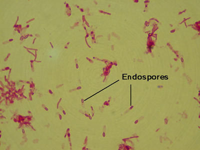
Oct 28, 15 Acid fast stain of Echerichia coli 400x on a compound light micrscope Echerichia coli is a gram negative, non spore forming capsulated bacteriaSUMMARY The shape of Escherichia coli is strikingly simple compared to those of higher eukaryotes In fact, the end result of E coli morphogenesis is a cylindrical tube with hemispherical caps It is argued that physical principles affect biological forms In this view, genes code for products that contribute to the production of suitable structures for physical factors to act uponMalachite green primary staining step of endopore stain with slide being heated over water bath;


Solved In This Section You Will View A Random Set Of Micr Chegg Com


Science Source Stock Photos Video Escherichia Coli And Staph Aureus
4 Applying counterstain (safrinin) to bacterial smear as last step of endospore stain;After Gram staining, E coli are pink in color This result is interpreted as _____ E primary stain C The function of a mordant in staining procedures is to C 400X D 4000X C Why is a specimen smaller than 0 nm not visible with a light microscope?E Coli Gram Stain And Cell Morphology E Coli Micrograph Gram Stain Demonstration Slide 400x 2 A Slide Demonstrati Flickr S Epidermidis Gram Stain Escherichia Coli Free Images Public Domain Images Pbstatemicrobiology Licensed For Non Commercial Use Only Gram


Biol 2


Micromorphology Slides Microbiology Resource Center Truckee Meadows Community College
Instead, their genetic material floats uncoveredEndospore stained slide, with control Bacillus on left, negative control E coli on right, and unkown in centerFigure 3 In this specimen, the grampositive bacterium Staphylococcus aureus retains crystal violet dye even after the decolorizing agent is added Gramnegative Escherichia coli, the most common Gram stain qualitycontrol bacterium, is decolorized, and is only visible after the addition of the pink counterstain safranin


Solved In This Section You Will View A Random Set Of Micr Chegg Com
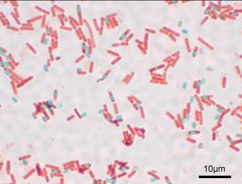


Print Micro Exam 2 Flashcards Easy Notecards
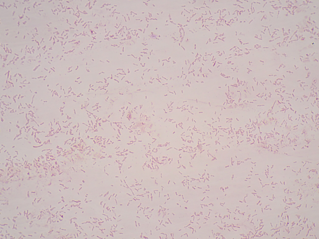
Diffusely adherent E coli (DAEC) Gram Stain E coli is described as a Gramnegative bacterium This is because they stain negative using the Gram stain The Gram stain is a differential technique that is commonly used for the purposes of classifying bacteria The staining technique distinguishes between two main types of bacteria (gramIntroduction Escherichia coli is a rod‐shaped, Gram‐negative bacterium, and classified as a member of the family Enterobacteriaceae within the Gammaproteobacteria classEscherichia coli is among one of the well‐studied bacteriaEscherichia coli can grow rapidly under optimal growth conditions, replicating in ~ min Many gene manipulation systems have been developed using E coli asE Coli E Coli under the microscope at 400x E Coli (Escherichia Coli) is a gramnegative, rodshaped bacterium Most E Coli strains are harmless, but some serotypes can cause food poisoning in their hosts The harmless strains are part of the normal flora of the gut Learn more about E Coli here Helicobacter Pylori


Www Mccc Edu Hilkerd Documents Bio1lab3 Exp 4 000 Pdf


The World S Best Photos Of Prokaryote Flickr Hive Mind Prokaryotes Microbiology Microbiology Study
Gram Staining Now that the slide has been heat fixed, you may now begin to stain the organism This is started by applying a few drops of crystal violet for 30 seconds, then rinsing with water for 5 seconds Next, cover the slide with Gram's Iodine and let sit for a full minute before rinsing with water for another 5 secondsA Gram positive B Gram negative If you gram stain sporeforming bacteria, you see many rod shaped, dark purple colored bacteria However, some of them have what appears to be a clear colored hole in the middle of the bacterialEscherichia coli (/ ˌ ɛ ʃ ə ˈ r ɪ k i ə ˈ k oʊ l aɪ /), also known as E coli (/ ˌ iː ˈ k oʊ l aɪ /), is a Gramnegative, facultative anaerobic, rodshaped, coliform bacterium of the genus Escherichia that is commonly found in the lower intestine of warmblooded organisms (endotherms) Most E coli strains are harmless, but some serotypes (EPEC, ETEC etc) can cause serious food
.jpg)


Biol 2


Gram Stain Demonstration Slide 400x 2 Marc Perkins Photography
Biology Of E Coli E coli (Escherichia coli) are a small, Gramnegative species of bacteriaMost strains of E coli are rodshaped and measure about μm long and 0210 μm in diameterThey typically have a cell volume of 0607 μm, most of which is filled by the cytoplasm Since it is a prokaryote, E coli don't have nuclei;Using Gram Staining (Lab 4L) (Cat # BE2) and in sharp focus on 100X before going to 400X, and center and focused on 400X before adding the immersion oil and going to 1000X Students often use too much light when using a microscope Decreasing the diaphragm E coli cells are Gram4 Applying counterstain (safrinin) to bacterial smear as last step of endospore stain;


52 Microbiology Unknown Project Cscc Bio 2215 Ideas Microbiology Medical Laboratory Medical Laboratory Science


Link Springer Com Content Pdf 10 1007 2f978 3 319 2 Pdf
Gram stain (CDC) Salmonella Nomenclature The genus Salmonella is a member of the family Enterobacteriaceae, It is composed of bacteria related to each other both phenotypically and genotypically Salmonella DNA base composition is 5052 mol% GC, similar to that of Escherichia, Shigella, and CitrobacterThe key difference between E coli and Pseudomonas aeruginosa is that E coli is a facultative anaerobic bacterial species that belongs to family Enterobacteriaceae and genus Escherichia, while P aeruginosa is an aerobic bacterial species that belongs to family Pseudomonadadaceae and genus Pseudomonas Both E coli and Pseudomonas aeruginosa are gramnegative, rodshaped and motile bacteriaWhich factor will NOT adversely affect the Gram reaction of a bacterium?


Escherichia Coli Light Microscopy


Biol 230 Lab Manual Lab 1
The key difference between E coli and Pseudomonas aeruginosa is that E coli is a facultative anaerobic bacterial species that belongs to family Enterobacteriaceae and genus Escherichia, while P aeruginosa is an aerobic bacterial species that belongs to family Pseudomonadadaceae and genus Pseudomonas Both E coli and Pseudomonas aeruginosa are gramnegative, rodshaped and motile bacteriaTHE PROCEDURE done individually We have cultures of E coli and Bacillus for you to gram stainThis will give you gram and gram – controls to check your procedure against You can use 2 slides, 1 for each bacterium, or you can divide one slide in half and smear each bacterium on the divided slideStaphylococci (staphylococcus aureus)gram positive, spherical bacteris that have developed antibiotic resistant strains, 400x gram stain stock pictures, royaltyfree photos & images microscopic view of bacterial pneumonia bacterial pneumonia is a type of pneumonia caused by bacterial infection gram stain stock illustrations


Bacterial Morphotypes In Sputum Gram Stain 100 Oil Immersion Field Download Scientific Diagram


Science Source Stock Photos Video Escherichia Coli And Staph Aureus
Enterobacter spp, Escherichia coli, Gram stain results, organism, likelihood that the culture was contaminated based on the organisms that are isolated, number of organisms that grow, and patient gender Susceptibility Testing ;Biology Of E Coli E coli (Escherichia coli) are a small, Gramnegative species of bacteriaMost strains of E coli are rodshaped and measure about μm long and 0210 μm in diameterThey typically have a cell volume of 0607 μm, most of which is filled by the cytoplasm Since it is a prokaryote, E coli don't have nuclei;THE PROCEDURE done individually We have cultures of E coli and Bacillus for you to gram stainThis will give you gram and gram – controls to check your procedure against You can use 2 slides, 1 for each bacterium, or you can divide one slide in half and smear each bacterium on the divided slide


Team Hkust Hong Kong Characterization 12 Igem Org


Q Tbn And9gcto 2gsvobxf2sagxghjthuqeairs5hk Ged4ppdotzlayzehab Usqp Cau
Instead, their genetic material floats uncoveredAfter Gram staining, E coli are pink in color This result is interpreted as _____ Gram negative Put the following steps of bacterial specimen preparation and staining in order a fixation b application of staining dyes c smear preparation 400X Why is a specimen smaller than 0 nm not visible with a light microscope?C 400X D 1000X E unable to be determined C 400X Is Escherichia coli Gram positive or Gram negative?


The World S Best Photos Of Prokaryote Flickr Hive Mind Prokaryotes Microbiology World Best Photos
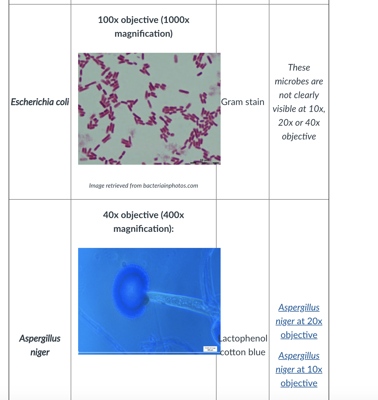


Please Answer From Penicilium To The End Thanks Fi Chegg Com
In order to perform a gram stain, your doctor will need to collect a sample of body fluid or tissue for analysis Their collection methods will vary depending on the type of sample they needEndospore stained slide, with control Bacillus on left, negative control E coli on right, and unkown in centerE Coli Gram Stain And Cell Morphology E Coli Micrograph Gram Stain Demonstration Slide 400x 2 A Slide Demonstrati Flickr S Epidermidis Gram Stain Escherichia Coli Free Images Public Domain Images Pbstatemicrobiology Licensed For Non Commercial Use Only Gram
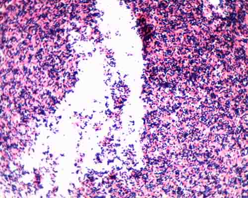


Gram Stain Microbiology Images Photographs
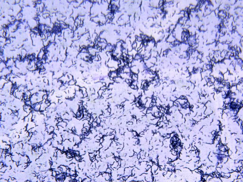


Solved In This Section You Will View A Random Set Of Micr Chegg Com
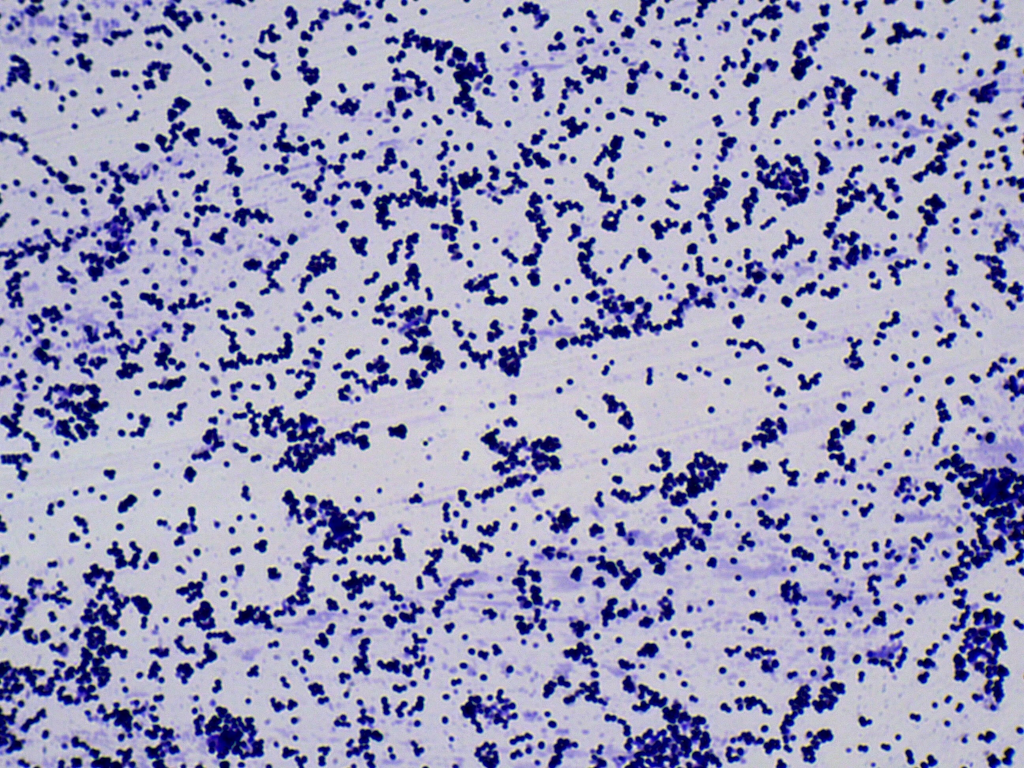
Gram stain demonstration slide, 400x 2 A slide demonstrating the gram stain On the slide are two species of bacteria, one of which is a gram positive coccus (Staphylococcus aureus, stained dark purple) and the other a gramnegative bacillus (Escherichia coli, stained pink) Seen at approximately 400x magnificationE Coli Bacteria Gram Negative Bacilli Gram Stain Lm X400 Escherichia Coli Bacteria E Coli Stock Footage Video 100 Electrically Induced Bacterial Membrane Potential Dynamics Surface Enhanced Raman Spectroscopy Of Microorganisms LimitationsEscherichia coli Gram Stain Gram negative bacilli fermenter;


Www Mccc Edu Hilkerd Documents Bio1lab3 Exp 4 000 Pdf


Microbiology Lablogatory
Gram stain (CDC) Salmonella Nomenclature The genus Salmonella is a member of the family Enterobacteriaceae, It is composed of bacteria related to each other both phenotypically and genotypically Salmonella DNA base composition is 5052 mol% GC, similar to that of Escherichia, Shigella, and CitrobacterEscherichia coli (/ ˌ ɛ ʃ ə ˈ r ɪ k i ə ˈ k oʊ l aɪ /), also known as E coli (/ ˌ iː ˈ k oʊ l aɪ /), is a Gramnegative, facultative anaerobic, rodshaped, coliform bacterium of the genus Escherichia that is commonly found in the lower intestine of warmblooded organisms (endotherms) Most E coli strains are harmless, but some serotypes (EPEC, ETEC etc) can cause serious foodEscherichia coli O157H7 was first identified as a human pathogen in 19 in the United States of America, following an outbreak of bloody diarrhea associated with contaminated hamburger meat In 06, there was an outbreak involving raw spinach, with 199 illnesses, 102 hospitalizations, 31 hemolytic uremic syndrome, a severe kidney condition


Ehrlich Staining A Simple Off The Shelf Diy Bio Stain For Bacteria Microorganisms Dna And Human Nuclei Exploring The Invisible
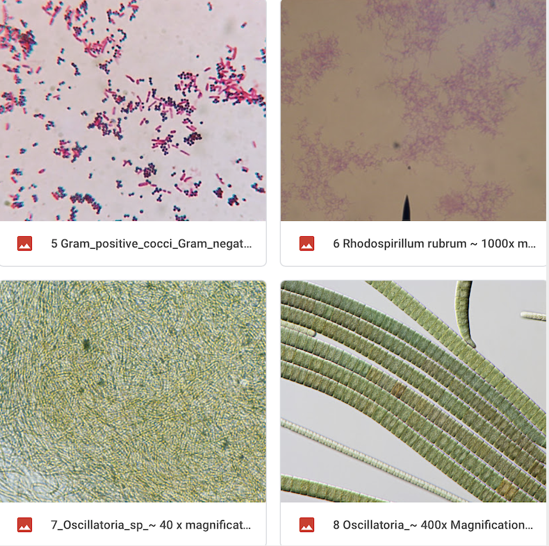


Solved Look At The 2 Gram Stain Slide This Is A Gram Sta Chegg Com
A coculture of E coli and Staphylococcus stained with DAPI (blue) and CF®633 WGA (red) Sphericallyshaped grampositive Staphylococcus stain with WGA, while rodshaped gramnegative E coli do not stain DAPI stains all cells blueMagnification 1000× Gram stain Result Gramnegative rods wwwbacteriainphotoscomMagnification 1000× Gram stain Result Gramnegative rods wwwbacteriainphotoscom


Gram Stain


Bacteria Laboratory Notes For Bio 1003 Bio 16 And Bio 3001
THE PROCEDURE done individually We have cultures of E coli and Bacillus for you to gram stainThis will give you gram and gram – controls to check your procedure against You can use 2 slides, 1 for each bacterium, or you can divide one slide in half and smear each bacterium on the divided slideBacteria E coli Must observe under 400x Very small & motile Gram Stain Staph Gram positive E coli Gram negative cocci in clusters rods (no arrangement) Refer to Lab Manual for directions Instructor to


Biol 2


Microbiology Page 21 Lablogatory


Microscope World Blog June 15
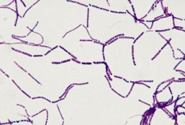


Print Micro Exam 2 Flashcards Easy Notecards


Microbiology Lablogatory


What Does An E Coli Bacteria Look Like Under A Microscope Quora


Gram Stain Demonstration Slide 400x 1 Marc Perkins Photography


Microscopy Gram Staining Microscope World Blog
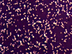


Welcome To Microbugz Negative Stain
.jpg)


Bacteria 1000x Gram Stain Bacteria 1000x Gram Stain Manufacturers Bacteria 1000x Gram Stain Suppliers Bacteria 1000x Gram Stain Exporters Bacteria 1000x Gram Stain In India


Gram Stain
.jpg)


Biol 2


Light Microscopy Streaked Images
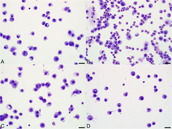


Pulmonary And Systemic Responses To Aerosolized Lysate Of Staphylococcus Aureus And Escherichia Coli In Calves Bmc Veterinary Research Full Text


Light Microscopy Streaked Images
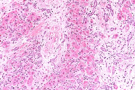


Afip Wsc 96 97 Conference 1


What Does An E Coli Bacteria Look Like Under A Microscope Quora


Qtelowidlcjkxm


1 276 Bacillus Subtilis Photos And Premium High Res Pictures Getty Images
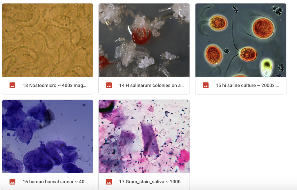


Solved Look At The 2 Gram Stain Slide This Is A Gram Sta Chegg Com
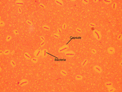


Micromorphology Slides Microbiology Resource Center Truckee Meadows Community College


Biol 2


Biol 230 Lab Manual Lab 1
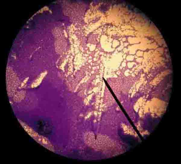


Gram Stain Microbiology Images Photographs


Q Tbn And9gcqkye60ou Johpr02n Mbv1fferrjpdh Lnct7ymdf5qhyia1ld Usqp Cau


Science Source Stock Photos Video Buccal Smear Lm


Www Mccc Edu Hilkerd Documents Bio1lab3 Exp 4 000 Pdf


Q Tbn And9gcqkye60ou Johpr02n Mbv1fferrjpdh Lnct7ymdf5qhyia1ld Usqp Cau
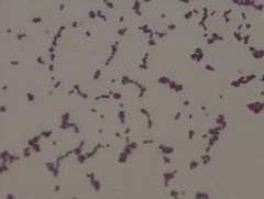


Bio 2 Identify Lab Slides Flashcards Cram Com


Micro Lab Final Flashcards Quizlet


Http Www Bio Rad Com Webroot Web Pdf Lse Literature a Pdf


Pin On E Coli


E Coli Negative Stain Page 1 Line 17qq Com


Q Tbn And9gctnncfjtcedl5fo0rq5mdljtvlng Qopeaabn2fkjwz27muvuqg Usqp Cau
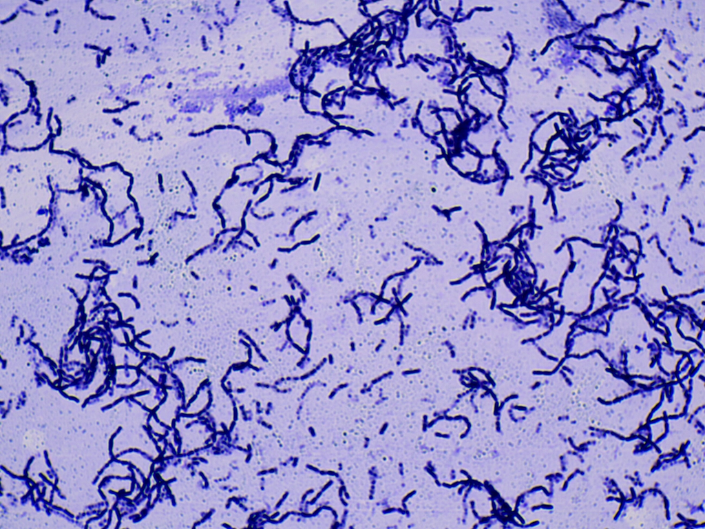


Solved In This Section You Will View A Random Set Of Micr Chegg Com


Biol 230 Lab Manual Lab 1
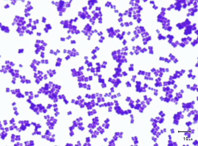


Print Micro Exam 2 Flashcards Easy Notecards


Www Scientistcindy Com Uploads 8 5 1 2 San Bernardino Valley College Micro Lab Manual 1 Pdf


Link Springer Com Content Pdf 10 1007 2f978 3 319 2 Pdf


52 Microbiology Unknown Project Cscc Bio 2215 Ideas Microbiology Medical Laboratory Medical Laboratory Science
.jpg)


Escherichia Coli 400x Escherichia Coli 400x Manufacturers Escherichia Coli 400x Suppliers Escherichia Coli 400x Exporters Escherichia Coli 400x In India


What Does An E Coli Bacteria Look Like Under A Microscope Quora
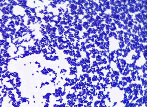


Bacterial Staining Microbiology Images Photographs And Videos Of Gram Acid Fast Endospore


E Coli Negative Stain Page 1 Line 17qq Com


Structure And Function Of Bacterial Cells


E Coli Slide Page 1 Line 17qq Com


E Coli Negative Stain Page 1 Line 17qq Com


347 Gram Stain Photos And Premium High Res Pictures Getty Images


Gram Stain


Gram Stain


Escherichia Coli Bacteria E Coli Stock Footage Video 100 Royalty Free Shutterstock


Www Mccc Edu Hilkerd Documents Bio1lab3 Exp 4 000 Pdf
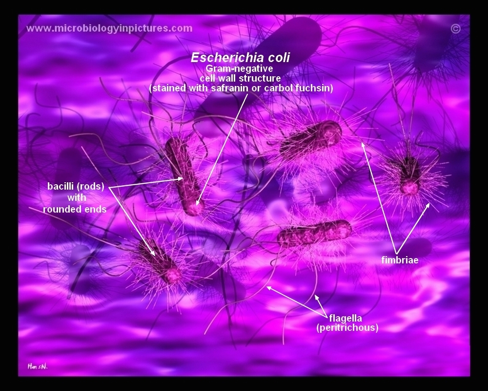


How E Coli Bacteria Look Like


Www Mccc Edu Hilkerd Documents Bio1lab3 Exp 4 000 Pdf


What Does An E Coli Bacteria Look Like Under A Microscope Quora


E Coli Negative Stain Page 1 Line 17qq Com


Microscope World Blog Microscopy Gram Staining


Photomicroscopy Photomicrography For Bacteriological And


Microbes Under The Microscope Flashcards Quizlet


Staphylococcus Aureus Under Microscope Microscopy Of Gram Positive Cocci Morphology And Microscopic Appearance Of Staphylococcus Aureus S Aureus Gram Stain And Colony Morphology On Agar Clinical Significance
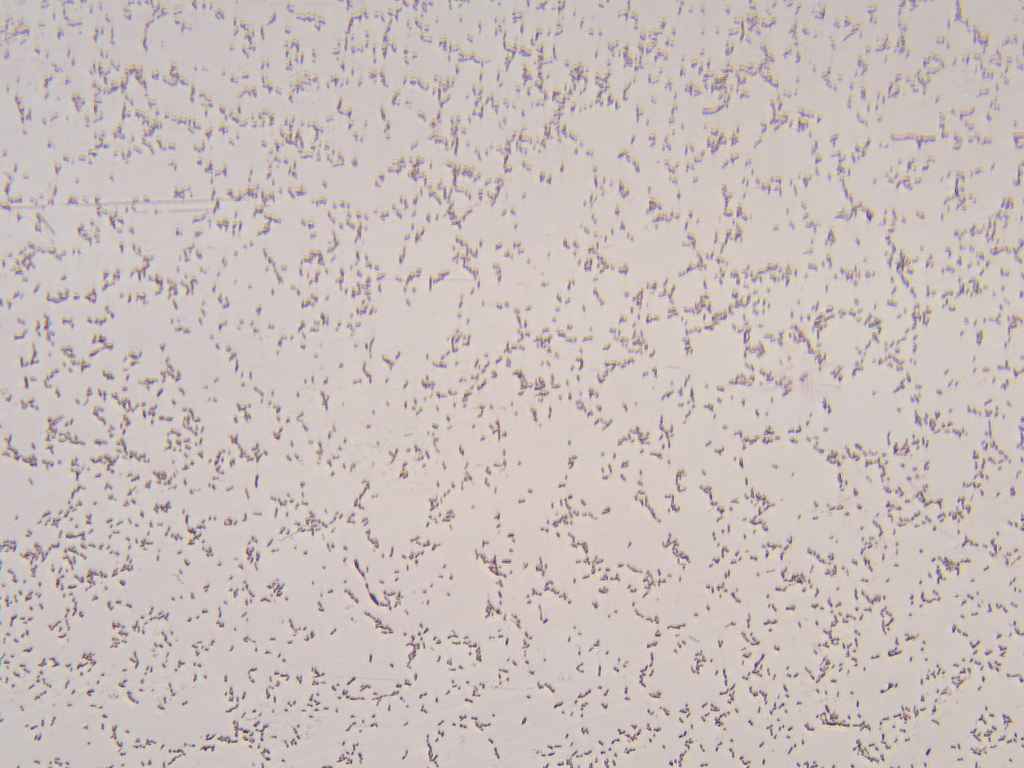


Solved In This Section You Will View A Random Set Of Micr Chegg Com
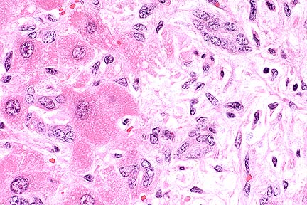


Afip Wsc 96 97 Conference 1
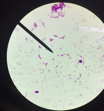


Print Micro Exam 2 Flashcards Easy Notecards


E Coli Gram Stain Introduction Principle Procedure And Result Interpret


Acridine Orange Staining Smear From Blood Culture Magnification Download Scientific Diagram


Biol 230 Lab Manual Lab 1
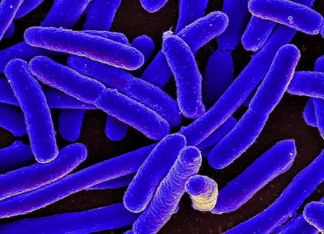


E Coli Under The Microscope Types Techniques Gram Stain Hanging Drop Method


Ehrlich Staining A Simple Off The Shelf Diy Bio Stain For Bacteria Microorganisms Dna And Human Nuclei Exploring The Invisible


Secreted Autotransporter Toxin Sat Induces Cell Damage During Enteroaggregative Escherichia Coli Infection


Biol 2


コメント
コメントを投稿